

What is Stargardt’s Disease (Fundus Flavimaculatus)

Stargardt’s Disease (Stargardt’s Macular Dystrophy) and its Treatment

History of Stargardt Disease

Stargardt's macular dystrophy was first described by German doctor Karl Stargardt in 1909. It is inherited in an autosomal recessive manner and is associated with mutations in the ATP Binding Cassette Retina-specific (ABCA4) gene.

The clinical symptoms of Stargardt's disease include bilateral central vision loss and discromatopsia, a type of color vision impairment. Fundoscopically, it is characterized by macular atrophy, beaten bronze macular lesions, and white-yellowish spots corresponding to the accumulation of lipofuscin at the level of the retinal pigment epithelium (RPE). The visual acuity can vary between 20/30 and 20/200.

Pathophysiology

Stargardt's disease is the most common form of juvenile-onset macular dystrophy. As mentioned before, it is caused by mutations in the ABCA4 gene, resulting in errors in the processing and transport of all-trans retinal in the photoreceptor visual cycle, leading to the formation of N- retinylidene-N-retinyl-ethanolamine (A2E) and the accumulation of lipofuscin in the photoreceptor outer segments and RPE layer.

The accumulation of lipofuscin is a key factor in causing the progressive bilateral central vision loss associated with Stargardt's disease. The disease is typically characterized by the presence of a "beaten bronze" or bull's eye appearance of the macula and subretinal lipofuscin spots.

Lipofuscin accumulation is influenced by various factors, including a deficiency in the conversion of all-trans retinol to 11-cis-retinal in RPE cells, as well as the phagocytosis of photoreceptor outer segments by RPE cells. The resulting formation of A2E can react with oxygen, leading to its conversion into A2E-epoxides upon exposure to light. A2E increases the sensitivity of RPE cells to blue light and can cause various toxic effects, including destabilization of mitochondrial and lysosomal membranes. Additionally, A2E can inhibit cytochrome c oxidase and disrupt electron flow in the respiratory chain, leading to energy metabolism deficiencies and the production of reactive oxygen species.
Stargardt Disease Sign, Symptoms and Diagnose

It has been shown that A2E accumulation and its toxic effects are light- dependent. However, destabilization of lysosomal and mitochondrial membranes can occur even in the absence of light. Studies have demonstrated that A2E can destabilize isolated mitochondria and lysosomes, indicating its ability to induce destabilization regardless of light exposure.
Alternatively, a study has proposed an alternative toxic pathway in RPE cells loaded with A2E. In this study, lysosomal destabilization was not observed, but the cells were unable to fully digest the phagocytosed photoreceptor outer segments within 24 hours. The accumulation of undigested phospholipids, which are a source of reactive oxygen species, was found to be a result of the circadian-regulated process of phagocytosis.
Image

Sign, Symptoms

The main symptom of Stargardt Disease is loss of visual acuity, which may range from 20/30 to 20/200  

  • Sensitivity to glare
  • Central vision loss ( center of retina and focus of vision is damaged ; leaving peripheral vision more intact ) : central vision loss is mostly responsible for the inability of reading by Stargardt Disease patients.Wavy vision, Blind spots, Bluriness, Impaired color vision,difficulty adapting to dim lighting, decreased color vision, loss of visual acuity
  • Strabismus (cross-eye) : cross-eye can occur by most of the patients with Stargardt Disease.
  • Depth perception failure : patients may have difficulties in discrimination of two objects that have a certain distance between each other. Patients see these objects as if they are aligned.
  • Come and go vision : some patients with Stargardt are complaining about fast disappearing and reappearing of objects wearing sunglasses to protect themselves from sunlight
  • Mothers with Stargardt Disease often complain about inability to keeping any eye contact with their children’s. 
Stargardt's disease typically affects young patients but can also present in adulthood and may be misdiagnosed as age-related macular degeneration (AMD).
Image

Stargardt's Disease Diagnose

The diagnosis of Stargardt's disease relies on a combination of medical history, epidemiology, angiography, autofluorescence (AF), optical coherence tomography (OCT), multifocal electroretinography (mfERG), visual field testing,gene analysis and full-field electroretinography (ffERG). Angiography typically shows a "choroidal silence" due to lipofuscin accumulation in the RPE, and hyperfluorescent spots may also be observed.

Genetics:

STGD1: The most common form of Stargardt disease is the recessive form
caused by mutations in the ABCA4 gene.

STGD3: There is also a rare dominant form of Stargardt disease caused by
mutations in the ELOVL4 gene.

STGD4: Associated with PROM1.

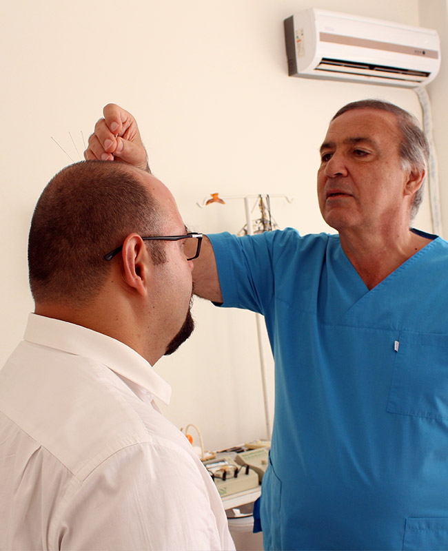
Treatment of Stargardt Disease with Electro-Acupuncture Method of Dr.Osman FIRATLI in Firatli Clinic

Treatment of Stargardt Disease with Electro-Acupuncture Method of Dr.Osman FIRATLI in Firatli Clinic

We offer Electro-Acupuncture treatment for patients with Stargardt Disease . Our treatment has shown its effectiveness of regaining the photoreceptors functions with more than 1000 patients with Stargardt Disease ; more than 5000 patients Retinitis Pigmentosa and Stargardt Patients.

The treatment and its success has been presented in several World-Congresses since 2007 with the medical evidences of our own patients; comparing their own visual field tests just before the treatment and after the treatment to indicate the improvement on both peripheral and central vision.

On this web-site you can view some videos of their own; presenting their own vision field test compare and how they benefited from our treatment.

Success Rate

In conventional medicine researches continue with the aim of preventing the disease from advancing further. That will even be considered as a huge success.

The aim of our treatment is not only preventing further deterioration of the eyesight but also improving the visual acuity and peripheral vision of our patients. With this purpose, our patients perform several medical tests at the eye hospitals before the therapy. These medical tests are repeated after the treatment to show the improvements that our patients also realize during their treatment period.

Considering that we only think our treatment is successful when the examinations at our clinic and the medical tests performed before and after clearly show the improvements on the eyesight and visual acuity.

Success rate of the therapy with Dr.Firatli Method is more than 90%
The combinations used during the therapy , applied treatment method and the electro-stimulation combinations depending on the type of the patient are unique and important factors on the high success rate.
Image
Fıratlı Clinic İstanbul

Follow Us

  Disclaimer: The testimonies and the developments published on this web-site vary from patient to patient. We can not take responsibility as Firatli Clinic that these developments apply to every patient...
© Copyright 2026 Fıratlı Clinic. All Rights Reserved.
Fıratlı Clinic Logo
Address

Address

Ataköy 1. KısımF26 Blok, Kat 5, Daire : 9Bakırköy / İstanbul
Fax

Fax

+90 212 661 0209
Sent Message Click To Call